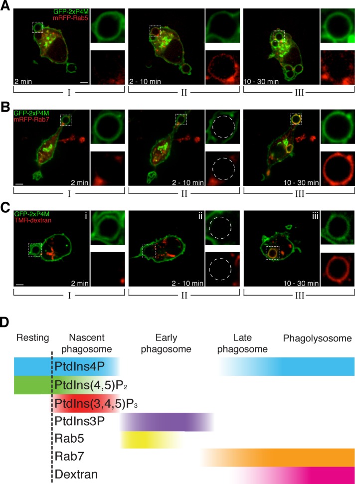
FIGURE 3:
Late phagosomes and phagolysosomes contain PtdIns4P. (A) Time-lapse gallery of confocal micrographs of RAW264.7 cells transiently coexpressing GFP-2xP4M and mRFP-Rab5 during phagocytosis of IgG-SRBCs. (B) Confocal micrographs of RAW264.7 cells transiently coexpressing GFP-2xP4M and mRFP-Rab7 during the course of phagocytosis of IgG-SRBCs. (C) Confocal micrographs of cells expressing GFP-2xP4M and exposed to TMR-labeled 10-kDa dextran for 3 h and chased for 30 min to label lysosomes. Images are representative of the distribution noted during the indicated time intervals after the onset of phagocytosis. Insets, magnifications of the area delimited by dotted white boxes. Scale bars, 5 μm. (D) Relative timing of the acquisition by phagosomes of different phosphoinositides and canonical markers of phagosome maturation. Qualitative only, intended to illustrate the relative order of events.

