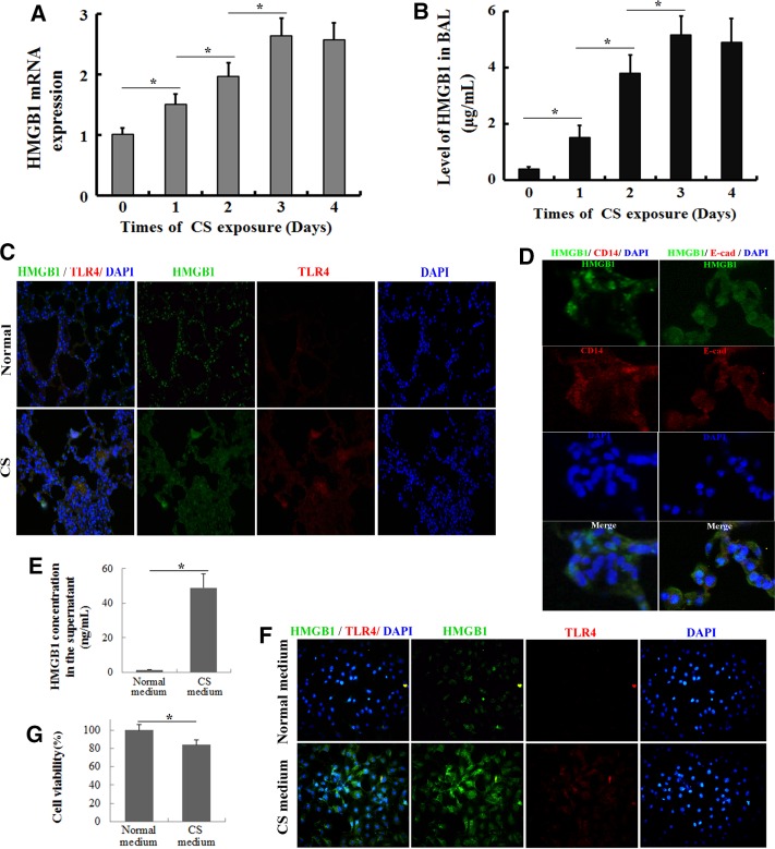
FIGURE 3:
HMGB1 translocation and release in the lung after CS exposure. The lungs and BAL fluid were obtained from C57BL/6J mice that had been exposed to the smoke from five cigarettes four times per day for various times (0, 1, 2, 3, and 4 d). (A) HMGB1 mRNA expression levels were determined by quantitative real-time PCR. (B) The concentrations of HMGB1 in the BAL fluid were determined by ELISA. Values are expressed as means ± SD from three independent experiments. *p < 0.05. (C) Double immunohistochemical staining for HMGB1 and TLR4 in the lung tissue after a 3-d CS exposure. (D) Double immunohistochemical staining for HMGB1 and CD14 (a marker of inflammatory cells) or E-cadherin (a marker of epithelial cells) in the lung tissue after a 3-d CS exposure. After MTE cells were stimulated with the CS-containing medium for 24 h, the HMGB1 content in the supernatant and HMGB1 translocation were determined by (E) ELISA and (F) immunofluorescence, respectively. (G) Cell viability was determined as a percentage of the levels in the nontreated control cells using the MTT assay. The results are displayed as means ± SD. from three independent experiments. *p < 0.05.

