
FIGURE 1:
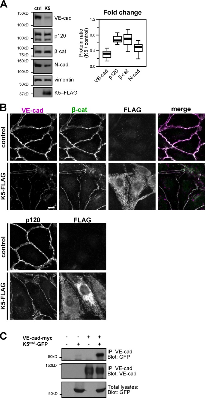
K5 down-regulates VE-cadherin. (A) K5-FLAG was expressed in cultures of the endothelial cell line HMEC-1 using adenoviral transduction. Control cells were uninfected. After 48 h, cells were harvested and the lysates analyzed by Western blot. Thick line, median band intensity; boxes, interquartile range; whiskers, 90% range (n = 7 sample pairs per protein); p < 0.01, VE-cadherin compared with p120; p < 0.05, VE-cadherin compared with β-catenin. (B) FLAG-tagged K5 was expressed in primary cultures of dermal microvascular endothelial cells. After 48 h, cells were fixed and stained for VE-cadherin, β-catenin, or FLAG (top) or p120 and FLAG (bottom). Bars, 10 μm. (C) VE-cadherin forms a biochemical complex with K5 RING mutant. VE-cadherin-myc and a ligase-dead RING mutant of K5-GFP were expressed in COS-7 cells as indicated. After 24 h, total cell lysates were immunoprecipitated with anti-VE-cadherin antibody, and the coprecipitation of mutant K5-GFP was analyzed by Western blot.