
FIGURE 4:
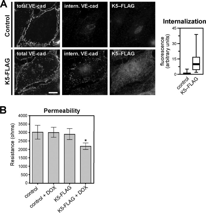
K5 induces VE-cadherin endocytosis and increased endothelial permeability. (A) K5-FLAG was expressed in primary dermal microvascular endothelial cells by adenoviral transduction. Cells were treated with 1 μg/ml doxycycline to suppress K5 expression until 6 h before beginning the assay. At 24 h after transduction, VE-cadherin endocytosis was measured using a fluorescence-based internalization assay. Internalized VE-cadherin (center column) was identified by antibody-labeling cell-surface VE-cadherin, incubating cells for 10 min to allow endocytosis, and then washing cells with a low-pH buffer to remove any antibody remaining on the cell surface. A second antibody was used to label the total VE-cadherin pool for comparison (left column). Thick line, median; boxes, interquartile range; whiskers, 90% range (n = 55–58 cells/group); p < 0.001. Bar, 20 μm. (B) Barrier function HDMECs expressing doxycycline-inducible K5-FLAG or control HDMECs was measured using an ECIS assay 48 h after K5-FLAG induction. Error bars represent SD (n = 3 control HDMEC and 21 K5-FLAG expressing HDMEC replicates); *p < 0.05 for K5-FLAG plus doxycycline compared with K5-FLAG control