
FIGURE 5:
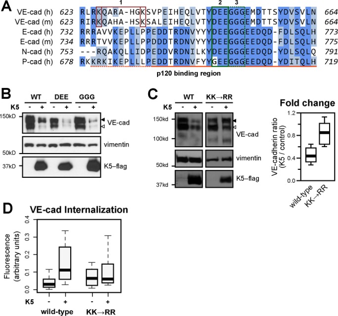
K5 induces VE-cadherin down-regulation through an alternate endocytic motif. (A) Multiple sequence alignment of the juxtamembrane domains of classical cadherins. 1, K626 and K633 mutated in C; 2, DEE646–648 mutated in B; 3, GGG649-651 mutated in B. The p120-binding region is marked with an orange line below the alignment: solid line, static binding region; dotted line, dynamic binding region. (B) Wild-type or mutant VE-cadherin–red fluorescent protein (RFP) and K5-FLAG were expressed in primary dermal microvascular endothelial cells by adenoviral transduction. VE-cadherin with a DEE646-648AAA mutation (DEE) does not bind p120 but is resistant to constitutive endocytosis (Nanes et al., 2012). VE-cadherin with a GGG649-651AAA mutation (GGG) does not bind p120 and undergoes constitutive endocytosis normally. At 48 h after transduction, cells were harvested and the lysates analyzed by Western blot. Empty arrowhead, endogenous VE-cadherin; filled arrowhead, VE-cadherin–RFP. (C) Wild-type (WT) or mutant (K626R, K633R; KK→RR) VE-cadherin–RFP was stably expressed in HMEC-1 cells using lentiviral transduction. K5-FLAG was expressed by adenoviral transduction 48 h before cells were harvested, and the lysates were analyzed by Western blot. Empty arrowhead, endogenous VE-cadherin; filled arrowhead, VE-cadherin–RFP. Thick line, median; boxes, interquartile range; whiskers, 90% range (six or seven sample pairs/group); P < 0.05. (D) Wild-type or KK mutant VE-cadherin–RFP was expressed in COS-7 cells by transient transfection. K5-FLAG was expressed by adenoviral transduction 24 h before measurement of VE-cadherin endocytosis over a 30-min period using a fluorescence-based internalization assay. KK, VE-cadherin K626R K633R, the K5-resistant mutant; Thick lines, median; boxes, interquartile range; whiskers, 90% range (25–30 cells/group). WT plus K5 compared with WT, p = 0.006.