
FIGURE 7:
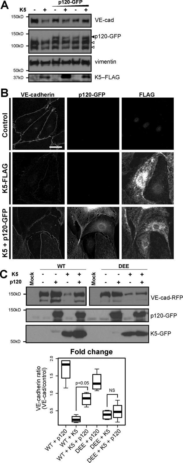
p120 binding protects VE-cadherin from down-regulation by K5. (A) K5-FLAG was expressed in primary dermal microvascular endothelial cells by adenoviral transduction. After 24 h, cells were additionally transduced to express varying levels of p120-GFP or transduced with an empty adenovirus as a control. After another 24 h, cells were lysed and analyzed by Western blot. Empty arrowheads, endogenous p120 isoforms; filled arrowhead, exogenous p120-GFP. (B) K5-FLAG or K5-FLAG and p120-GFP were expressed in primary dermal microvascular endothelial cells by adenoviral transduction. After 24 h, cells were fixed and processed for immunofluorescence. Bar, 20 μm. (C) p120 fails to protect the DEE mutant from K5-mediated down-regulation. Wild-type or DEE mutant VE-cad-RFP was expressed with K5-GFP in COS-7 cells by transient transfection as indicated. p120-GFP was expressed by adenoviral transduction. After 24 h, cells were lysed and analyzed by Western blot. For VE-cadherin (top), the right sample (DEE) was exposed longer than the left one (WT) because of decreased expression of the DEE mutant compared with wild type in COS-7 cells. Thick line, median; boxes, interquartile range; whiskers, 90% range (four sample pairs/group). Cadherin levels for cells expressing K5, p120, or K5 plus p120 were normalized to control cells expressing only WT cadherin or the DEE mutant, respectively. WT plus K5 plus p120 vs. WT plus K5: p < 0.05. DEE plus K5 plus p120 vs. DEE plus K5: not significant.