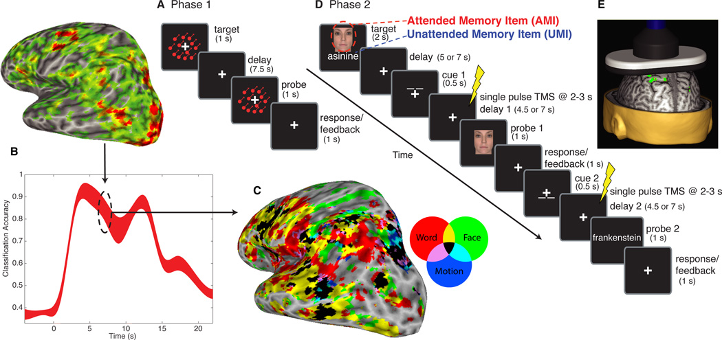
Fig. 1. General procedure.
In Phase 1, functional magnetic resonance imaging (fMRI) data were acquired while participants performed a one-item delayed-recognition task for words, faces, or directions of motion (A), and used for multivariate pattern analysis (MVPA). Classifiers trained on the delay-period (B) were used for subsequent analyses. For Experiment 1, these classifiers were used to decode fMRI activity from Phase 2 (Fig. 2). For Experiments 2 and 3, they were used in a whole-brain searchlight, conjunction-analysis to generate subject-specific maps of category-sensitive areas (C); non-overlapping areas were used for transcranial magnetic stimulation (TMS) targeting in Phase 2 (D). In Phase 2, single pulses of TMS (E) were delivered during the post-cue delay periods.

