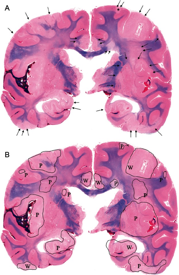
Fig. 2.

A, B - Case 1: Representative macroscopic coronal section of the cerebrum. Multiple infarcts with perifocal edema are scattered throughout the cortex and subcortical white matter (arrow). Note the flattened gyri with an unclear corticomedullary junction (Fig. 2A). Infarcts are demonstrated in black-edged area, P as patchy-shaped pattern and W as wedge-shaped pattern, respectively (Fig. 2B).
