
Fig. 4.
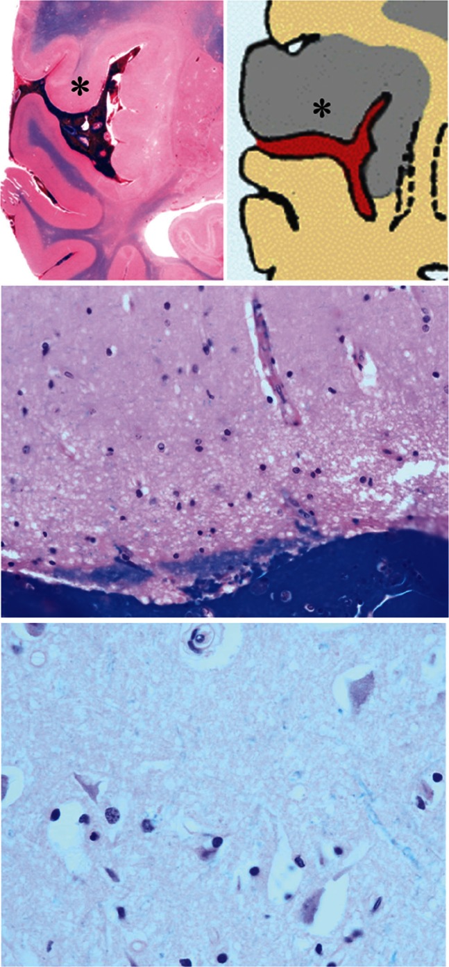
Case 1: Upper left: Macroscopic section. Note the unclear corticomedullary junction in the area with gyral swelling (*). Upper and lower right: Microscopic section of the area (*) where the subarachnoid clot tightly adhered to the pia mater. Note multiple cytotoxic edema and neuronal death indicative of acute ischemic changes.