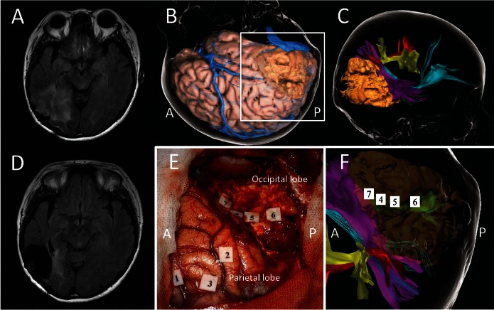
Fig. 3.
A case of recurrent low-grade glioma in the right temporal lobe. A: Preoperative FLAIR axial MRI showing a hyper lesion in right temporo-occipital area. B: Three-dimensional cortical image in operative view. C: Tractography with a recurrent tumor (beige); the inferior fronto-occipital fascicle (cyan), the superior longitudinal fascicle II (red) and III (yellow), and middle longitudinal fascicle (purple). D: Postoperative FLAIR axial MRI. E and F show an intraoperative photo and preoperative tractography within the white square in B. E: An intraoperative photo taken after surgical resection of tumor with preservation of positive mapping areas elicited by cortical and subcortical electrical stimulations. Cortical mappings: Dysesthesia of left hand in the postcentral gyrus (tag 1), and rightward deviation in the line bisection task (tags 2 and 3). Subcortical mappings: phosphene in the left superior quadrant area (tags 4–6) and topographical disorientation (tag 7). F: Tags 4–6 in the subcortical positive mapping areas are overlapped on the optic radiation (green) in the preoperative tractography. A and P indicate the anterior and the posterior side, respectively.

