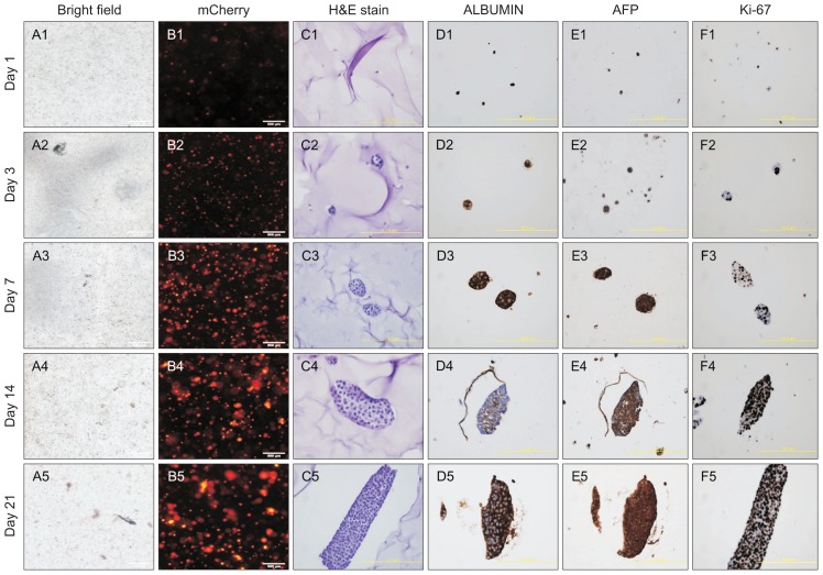
Fig. 3.
Proliferation of HepG2 cells in the three-dimensional (3D) culture system. (A1–5, B1–5) Bright field image of HepG2 cells grown in 3D culture (A1–5) and immunofluorescence image of HepG2 cells labeled with mCherry (B1–5). Bar, 500 μm. (C1–5, D1–5) Images of H&E staining (C1–5) and immunohistochemistry using antibodies against ALBUMIN (D1–5), AFP (E1–5), and Ki-67 (F1–5). Bar, 400 μm. The images were captured on days 1, 3, 7, 14, and 21 after seeding the HepG2 cells on 3D-printed scaffolds.

