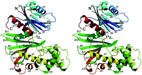
FIG. 1.
Ribbon model of the ecGlK monomer. Model uses rainbow colors from the N terminus (blue) to the C terminus (red). β-strands and α-helices are numbered sequentially from the N to C terminus. This and subsequent figures were prepared with either PyMOL (18; http://www.pymol.org) or Molscript (19) and Raster3D (41).

