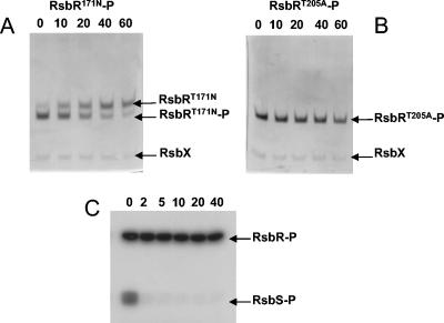
FIG. 5.
Dephosphorylation of RsbR-P by RsbX. (A) Dephosphorylation of 10 μM RsbRT171N-P-RsbS by 2.5 μM RsbX. At the time intervals (in minutes) indicated at the top of the gel, samples of the reaction mixture were subjected to alkaline urea-PAGE, and the gel was stained by Coomassie blue. (B) Dephosphorylation of 10 μM RsbRT205A-P-RsbS by 2.5 μM RsbX. Samples were treated as described for panel A. (C) After phosphorylation of the RsbR-RsbS complex by RsbT in the presence of 20 μCi of [γ-32P]ATP, a 10 μM concentration of the radiolabeled RsbR-RsbS complex was mixed with 2.5 μM RsbX. At the indicated time intervals (in minutes), reaction samples were mixed with 3× SDS loading buffer and heated for 3 min at 95°C. Samples were analyzed by SDS-13.5% PAGE and revealed by the use of a phosphor screen.

