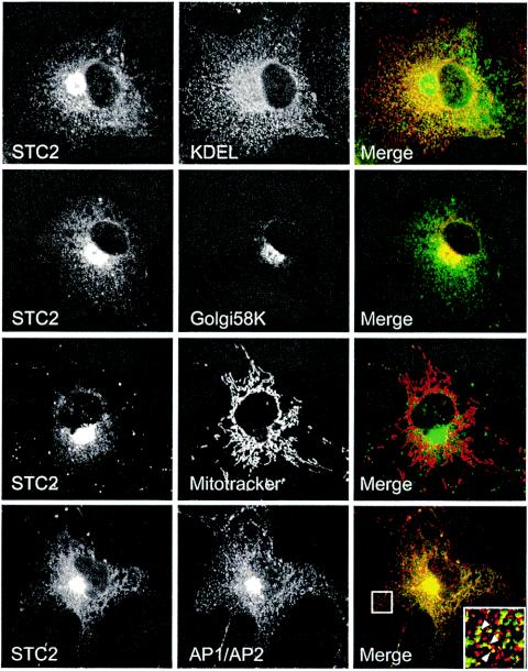
FIG. 4.
Immunofluorescence localization of STC2 to the ER and Golgi apparatus. COS cells transfected with STC2 were fixed with 4% paraformaldehyde, permeabilized in 0.2% Triton X-100, and double labeled with STC2 antiserum (green) and monoclonal antibodies (red) to KDEL (marker of ER), 58K (Golgi apparatus), and AP1/AP2 (TGN-derived secretory vesicles and plasma-membrane derived endocytic vesicles). Mitochondria were labeled by incubating cells with MitoTracker red. Images were acquired on a laser scanning confocal microscope. Note the extensive colocalization of STC2 with the ER, the Golgi/TGN, and the presence of STC2 in some AP1/AP2-positive small vesicles.

