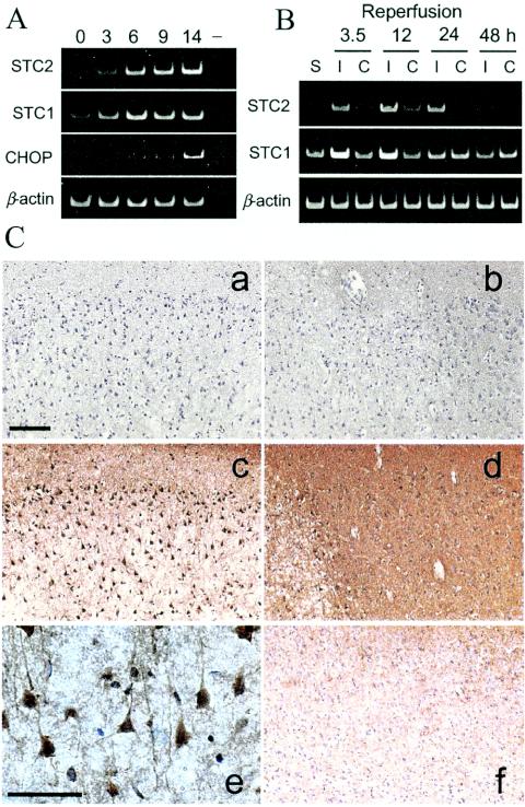
FIG. 8.
Induced expression of STC2 in ischemic rat brain. (A) Rat PC12 cells were subject to hypoxic conditions for the indicated periods. Levels of STC2, STC1, BiP, and β-actin mRNA were evaluated by RT-PCR with specific primers. Note the coordinated induction of STC2 and STC1 expression by hypoxia. (B) Rats were subject to transient MCA occlusion, and total RNA was extracted from cortex after reperfusion for the indicated periods. STC2, STC1, and β-actin mRNA levels were examined by RT-PCR. Lanes: S, sham-operated control; I, ischemic side cortex; C, contralateral side cortex. (C) Immunohistochemical analysis of STC2 expression in ischemic rats after 12 h of reperfusion. Sections were stained with preimmune serum (a and b) or STC2 antibody (c to f). Subpanels: a and c, ischemic core; b and d, penumbra; e, higher magnification of STC2 staining in ischemic cortex; f, contralateral cortex. Scale bar, 100 μm. Note the overall increase in STC2 immunoreactivity in the penumbra compared to the contralateral cortex and the intense staining of vulnerable neurons within the ischemic core.

