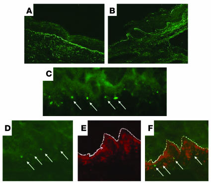
Figure 4.
IgA dermal deposit localization. (A) FITC staining identifies IgA deposition in perilesional uninvolved areas of NOD Abo DQ8+ mice with gluten-dependent blistering. Original magnification, ×20. (B) Deposition of IgA at the tips of papillae. Original magnification, ×20. (C) Granularity of the IgA deposits below the basement membrane zone. Original magnification, ×100. (D) FITC staining identifies IgA. Original magnification, ×100. (E) Rhodamine Red-X staining identifies collagen IV. Original magnification, ×100. (F) Composite picture with both IgA staining and collagen IV staining. Original magnification, ×100. Arrows identify IgA deposits, and the basement membrane zone is highlighted by a dotted white line.

