Figure 2.
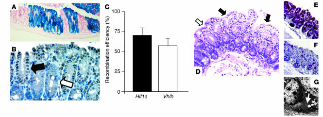
Fabp-Cre–mediated recombination in the colon: analysis of Fabp-Cre activity and histological characterization of mutant mice. (A and B) Fabp-Cre activity as determined by a lacZ reporter transgene and immunohistochemical staining for Cre-recombinase. (A) Section of the cecum stained with X-gal. Recombination occurs always in all cells of an individual crypt. Magnification, ×100. (B) Paraffin-embedded section of the colon from a Vhlh mutant mouse. Consistent with the recombination process in the lacZ transgenic mice, all cells within a Cre-positive crypt display nuclear-localized Cre signal (filled arrow), while all cells in a negative crypt stain negatively (open arrow). Magnification, ×400. (C) Determination of the efficiency of recombination by quantitative PCR. Levels of expression of the recombined gene (Hif1a or Vhlh) were compared with the expression levels of a non-recombining control gene in genomic DNA from isolated colonic epithelial cells (n = 3 for each genotype). (D–G) Clear-cell changes in the Vhlh mutant colon. (D) Luminal epithelial cells with pronounced clearing are indicated by filled arrows; the open arrow points to adjacent luminal cells without clearing. Clearing is a result of glycogen accumulation in Vhlh mutant cells, as shown by diastase-sensitive PAS staining. (E) PAS stain without diastase treatment. (F) PAS stain after diastase treatment of the adjacent colon. (G) Ultrastructural analysis of clear cells showed accumulation of cytoplasmic material that is morphologically consistent with glycogen (white arrow). The star indicates the nucleus of a clear cell. Magnification in D–F, ×200.

