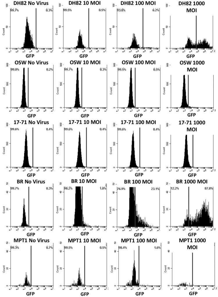
Fig 3. GFP Expression by canine hematopoietic origin cancer cells; Histiocytoma (DH82), Lymphoma (1771 and OSW) and Mast Cell Tumor (MPT1 and BR) cells at three different MOIs.
Cells were transduced by adenovirus Ad5G/L at three different multiplicity of infection (MOI 10, 100, and 1000 virus particles per cell). GFP expression in cells was analyzed by flow cytometry 48 hours after Ad5G/L infection.

