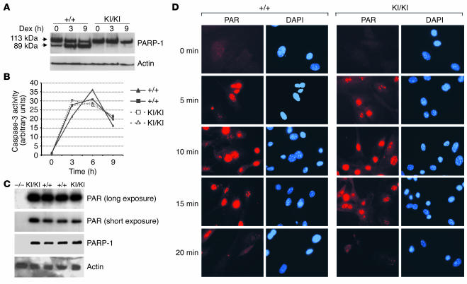
Figure 2.
Characterization of mutant PARP-1. (A) Western blot analysis showing the cleavage of PARP-1 in cell extracts from PARP-1+/+ and PARP-1KI/KI thymocytes treated with 1 μM dexamethasone (Dex) for 3 and 9 hours. Note intact PARP-1 in PARP-1KI/KI cell extracts. Actin was used as a loading control. This experiment was repeated at least 2 times. (B) Caspase-3 activity was measured using 2 sets of PARP-1+/+ and PARP-1KI/KI thymocytes after treatment with 1 μM dexamethasone for 3, 6, and 9 hours. (C) Enzymatic activity analysis in PARP-1KI/KI MEFs. Cell extracts from indicated genotypes were prepared and the polymer (PAR) formation was visualized by incubating the blot with 32P-labeled NAD+. The blot was re-hybridized with anti–PARP-1 and anti-actin antibodies. PARP-1–/– (–/–) MEFs were used as a control. This experiment was repeated at least 2 times. (D) Immunofluorescence staining of polymers (PAR). MEFs were incubated with 200 μM H2O2, fixed at the indicated time points, and labeled with the anti-PAR antibody (red). Nuclei were stained with DAPI (blue).

