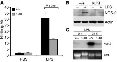
Figure 4.
Induction of NO and NOS-2 in PARP-1KI/KI macrophages by LPS. (A) PARP-1+/+ and PARP-1KI/KI macrophages were treated with LPS (1 μg/ml) for 24 hours, and nitrite in the medium was measured. The data are from pooled macrophages of 4 mice, values are the mean of duplicate measurements, and the study was repeated at least 3 times. (B) Western blot analysis of NOS-2 from macrophages treated with or without LPS for 24 hours. Note a great reduction of NOS-2 expression in PARP-1KI/KI cells compared with PARP-1+/+ samples. Actin was used as a loading control. This experiment was repeated three times. (C) Northern blot analysis of nos-2 expression in macrophages treated with LPS for 24 hours. Ethidium bromide staining of 28S rRNA was used as a loading control. Note a significant reduction in nos-2 expression in PARP-1KI/KI macrophages compared with PARP-1+/+ counterparts. This Northern blot is from 1 of the 2 experiments.

