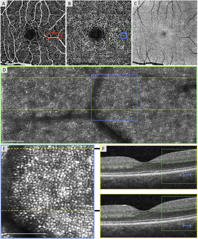
Fig 1. Normal Photoreceptors in an Area of Non-Flow of the Superficial Capillary Plexus (SCP).
Case 1, right eye. (A) Optical coherence tomography angiography (OCTA) of the SCP shows a relatively normal contour of the foveal avascular zone (FAZ) with focal areas of capillary non-flow inferior and superior to the FAZ, including an area imaged by adaptive optics scanning laser ophthalmoscopy (AOSLO) (red circle). (B) OCTA of the deep capillary plexus (DCP) with location of AOSLO montage (green box) and enlarged inset (blue box). DCP shows a normal FAZ, robust capillaries throughout, and a vessel density of 63.46%. (C) En face structural OCT image segmented at the inner segment / outer segment (IS/OS) and the outer segment / retinal pigment epithelium (OS/RPE) junctions is unable to resolve the photoreceptor mosaic. (D) AOSLO montage stitched from 2° x 2° images with location of B-scans (yellow lines) and enlarged inset below (blue box). (E) Enlarged 1° x 1° AOSLO image from montage with heterogeneity packing index of 0.432. Dotted lines indicate location of B-scans. (F) Spectral domain (SD)-OCT from the OCTA device showing robust IS/OS and OS/RPE bands. Green box and blue lines show location of AOSLO montage and enlarged inset, respectively. Green lines indicate the segmentation boundaries for the DCP. White scale bars in A, D and E are 100 μm.

