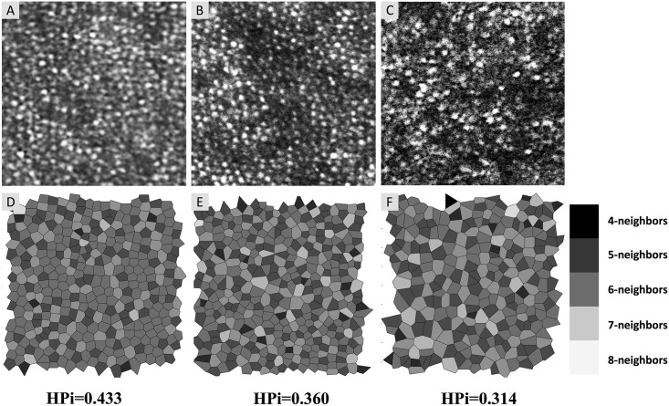
Fig 2. Adaptive Optics Scanning Laser Ophthalmoscopy (AOSLO) Images and Corresponding Voronoi Diagrams with Heterogeneity Packing Index (HPi).
(A-C) AOSLO images of cone mosaic (200 by 200 pixels taken from 1° x 1° images). (D-F) Voronoi tessellation corresponding to the image above with shading of cells indicating the number of neighboring photoreceptor cells, from dark (four neighbors) to light (eight neighbors). A lower HPi represents a larger deviation from the normal packing arrangement of cones. (A and D) Cones in an eye with DR without deep capillary plexus (DCP) non-flow (Case 3, HPi = 0.433). (B and E) Cones in an area of DCP non-flow (Case 10, HPi = 0.360). (C and F) Cones in an area of DCP non-flow (Case 8, HPi = 0.314).

