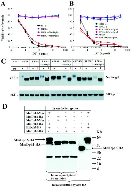
FIG. 3.
Complementation of the diphthamide-deficient CHO mutant cells by transfection with the respective mouse DPH genes. (A) Expression of MmDph1 in CG-4 PR303 cells complements their DT-resistant phenotype. Representative purified clones of PR303 cells transfected with mouse DPH1 or mouse DPH2 were incubated with various concentrations of DT for 48 h, and MTT was added to determine cell viability. Optical density readings were normalized to values for cells that received no toxin to calculate percent viability. (B) Expression of MmDph2 in CG-3 RPE33d cells and MmDph5 in CG-1 RPE3b cells complements their DT-resistant phenotypes. Cytotoxicity of DT to representative clones of RPE33d cells transfected with mouse DPH1 or mouse DPH2 and to a representative clone of RPE3b cells transfected with mouse DPH5 were determined as in panel A. (C) The CHO cell lysates were incubated with or without DT at room temperature for 30 min and then analyzed by either native PAGE (top) or SDS-PAGE (bottom), followed by Western blotting using an antibody against a linear peptide of the carboxyl terminus of eEF-2. +, present; −, absent. (D) Dph1 and Dph2 can physically interact in vivo. Combinations of HA- or Myc-tagged MmDph1, MmDph2, HsDph3, and MmDph5 expression plasmids as indicated were transiently transfected into CHO K1 cells for 24 h. Cell lysates were prepared and immunoprecipitated using an agarose-conjugated anti-Myc antibody, followed by Western blotting using an anti-HA antibody. Expression of MmDph5-HA in MmDph2-Myc- and MmDph5-HA-transfected cells was evidenced by direct Western blot analysis of the cell lysate using the anti-HA antibody. The two bands common in lanes 1 to 4 are to the heavy and light immunoglobulin G chains.

