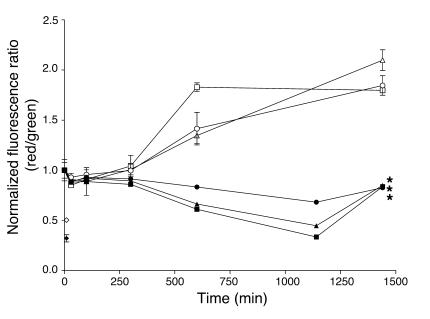
Figure 6.
Actinonin selectively causes mitochondrial membrane depolarization in a time- and dose-dependent manner. RL lymphoma cells (bottom 3 curves) were incubated with actinonin at 10 μg/ml (filled circles), 20 μg/ml (filled triangles), or 100 μg/ml (filled squares), treated with JC-1 dye, and analyzed by flow cytometry. Actinonin treatment led to a time- and dose-dependent depolarization of the mitochondrial membrane, as evidenced by the significant decrease (*P < 0.05) in the fluorescent red/green ratio. CCCP (filled diamond) was used as a positive control and showed depolarization after 10 minutes of treatment. Normal peripheral blood lymphocytes (top 3 curves) were incubated with actinonin at 10 μg/ml (open circles), 20 μg/ml (open triangles), or 100 μg/ml (open squares), treated with JC-1 dye, and analyzed by flow cytometry. There was no time- or dose-dependent depolarization of the mitochondrial membrane with actinonin treatment, although the CCCP control (open diamond) was effective at 10 minutes. All graphed values (mean ± SD) represent a normalized fluorescence ratio (red/green) calculated by division of the ratio for each time point by the ratio at time 0 (RL cells at t0, 1.853 ± 0.079; normal cells at t0, 0.349 ± 0.106).

