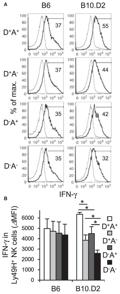
Figure 2. Ly49D and Ly49A Enhance Activation and IFN-γ Production of B10.D2 Ly49H+ NK Cells in the Early Course of MCMV Infection.

B6 mice were infected with 1 × 10⁴ pfu MCMV. B10.D2 mice were depleted of CD8+T cells onthe day before infection and theninfected with 5×10⁴ pfu MCMV.
(A) IFN-γ production of Ly49H+ NK cell subsets expressing Ly49D and or Ly49A in the spleen on day 1.5 pi. Data are representative of two experiments (n = 3–4 in each experiment).
(B) Delta mean fluorescent intensity (ΔMFI) of IFN-γ in Ly49H+ NK cells was quantified. Data were pooled from two experiments (n = 7 mice in each group). Bold and thin lines represent Ly49H+ NK cells in infected mice and Ly49H+ NK cells in uninfected mice, respectively. *p < 0.05. Error bars show SEM.
