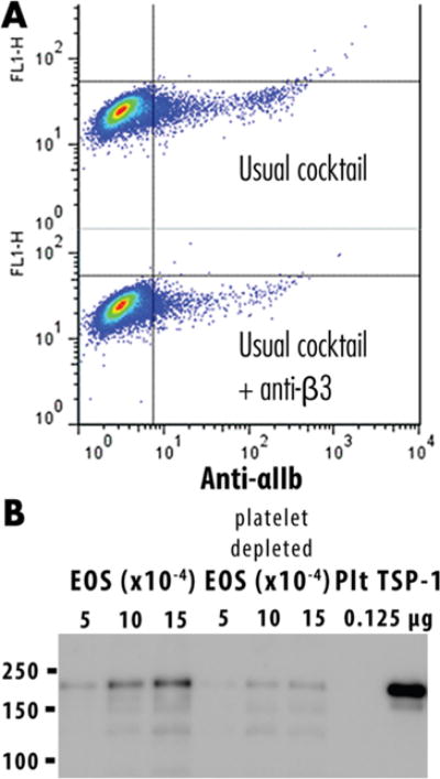
Figure 5.

Depletion of platelet contamination. (A) Addition of the anti-CD61 (β3 integrin) purification step yielded eosinophils that were less reactive with anti-CD41 αIIb integrin, the partner of β3 integrin, as shown by flow cytometry. (B) Immunoblotting demonstrates that the platelet-depleted eosinophils contain less THBS1.
