Figure 1.
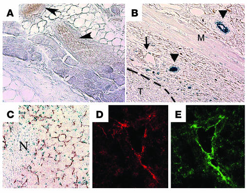
Induction of S1P1 expression in tumor xenografts. Normal skin tissue (A) and subcutaneous tumor nodules (B) from the back of S1p1+/–LacZ mice were excised and stained with X-gal to detect the expression of β-gal marker as described. Sections were cut and imaged under a bright-field microscope using a ×20 objective lens. β-gal expression (blue) was detected in blood vessels only in mice with the growing tumor (arrowheads in B). However, blood vessels in normal skin tissues (arrowheads in A) and few blood vessels with veinlike morphology in tumor-bearing tissue (arrow in B) were S1P1-negative. T, tumor; M, skeletal muscle layer. Dashed line indicates margin of the tumor. (C) X-gal–stained, fixed tissues were counterstained with anti–CD31 antibody to detect blood vessels, and the intratumor region is shown. At least 5 animals were used in this study. N, necrotic center. (D and E) Frozen sections of the subcutaneous implants of the tumor were analyzed in an immunofluorescence assay with the anti–S1P1 antibody (red) and anti–CD31 antibody (green) and imaged by a confocal microscope as described in Methods. Note that intratumoral blood vessels express S1P1, whereas tumor cells do not express this receptor.

