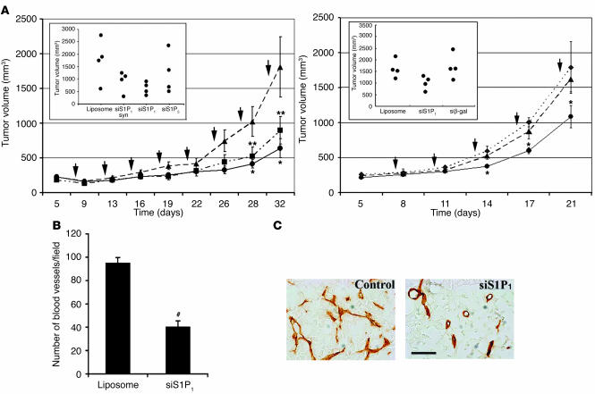
Figure 4.
Inhibition of tumor growth and angiogenesis by S1P1 siRNA. (A) Lewis lung carcinoma cells were implanted subcutaneously and allowed to establish as growing tumors, and various siRNA-liposome complexes were injected into the tumor every 3 days (where indicated by arrows) as described. Tumor volume was measured and plotted at various times following treatment with synthetic or multiplex siRNA for S1P1 (synthetic siS1P1, squares; multiplex siS1P1, circles) or liposome alone (triangles). The inset shows the tumor volume at 32 days for additional controls. Data represent mean ± SE of an experiment that was repeated twice. The right panel shows an independent experiment repeated with the inclusion of another control siRNA (multiplex β-gal siRNA, diamonds). n = 4; *P < 0.05; **P < 0.1. (B) Intratumoral microvessel density was quantified from multiple fields as described after CD31 staining. #P < 0.0003. (C) Morphology of tumor vessels is shown from a representative field. Note that vessel morphology and density are altered by S1P1, but not control, siRNA. Scale bar: 20 μm.

