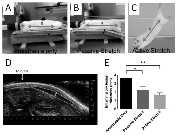
Figure 1. In vivo stretching and subacute inflammation.
Methods used for anesthesia only (A), passive stretch (B) and active stretch (C). The distance between shoulder and hip (arrows) is ~25% greater in both passive and active stretch, compared with anesthesia only. “X” indicates location of carrageenan injection. (D) Method of inflammatory lesion measurement on ultrasound images. The inflammatory lesion is outlined with a dashed line and the thickness measurement (double arrow) is taken perpendicular to the skin tangent at the point of maximum subcutaneous tissue thickness. (E) Two weeks after subcutaneous carrageenan injection, there was a reduction in inflammatory lesion thickness with both passive and active stretching, compared with anesthesia alone, (* p<0.05, **p<0.01, N=6 rats/group for active and passive stretch and N=5 rats for anesthesia alone). The difference between active and passive stretching was not statistically significant (p=0.27).

