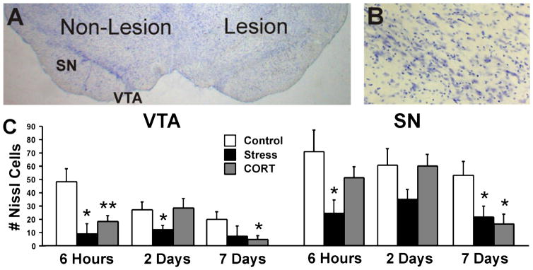
Fig. 9.
Nissl staining in VTA and SN. (A) Representative Nissl-stained section illustrating the non-lesion and lesion hemispheres. (B) Higher magnification of lesion substantia nigra (100×). (C) The number of Nissl-positive cells in VTA and SN in the lesion hemisphere. There was a significant acceleration of cell loss in stress-treated rats and reduced nigral cell numbers at 7 days post-lesion. Asterisks indicate significant differences: *P = 0.05; **P = 0.01; unpaired t-test compared with lesion control animals. VTA, ventral tegmental area; SN, substantia nigra pars compacta.

