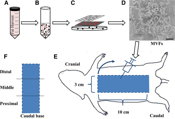
Fig. 1.

Schematic depicting the MVF isolation procedure and injection into the dorsal skin flap. A and B, Adipose tissue was harvested, minced, digested with collagenase, centrifuged (400g × 4 min), and (C) filtered through 500 μm and 30 μm filters to (D) isolate a heterogeneous mixture of MVFs. Black scale bar represents 100 μm. E, View of dorsal side of the rat with a diagram of the 10 × 3 cm skin flap. Dashed lines indicate edges that were cut and flap was raised (blue arrow) in a cranial to caudal fashion with the caudal side remaining intact. Injections were performed by injecting MVFs in 10 evenly dispersed sites in the distal half. Control animals underwent the identical surgery with sterile PBS injected alone as a control. F, Diagram of the 3 regions (distal, middle, and proximal) into which the flap was separated for perfusion and microvessel density measurements.
