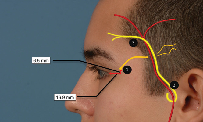
Fig. 3.

Temporal injection sites. Image showing the relationship of the AT (bright yellow), ZTBTN (dark yellow), and the anterior and posterior branches of the superficial temporal artery (red) to the temporal injection sites. The ZTBTN is on a different fascial plane than the AT and superficial temporal artery laterally. 1, ZTBTN injection site, which is commonly 1.5 cm behind the emergence of this nerve from the deep temporal fascia; 2, proximal AT corresponding to the fascial compression bands; 3, distal AT corresponding to the anterior temporal artery crossing the AT nerve.
