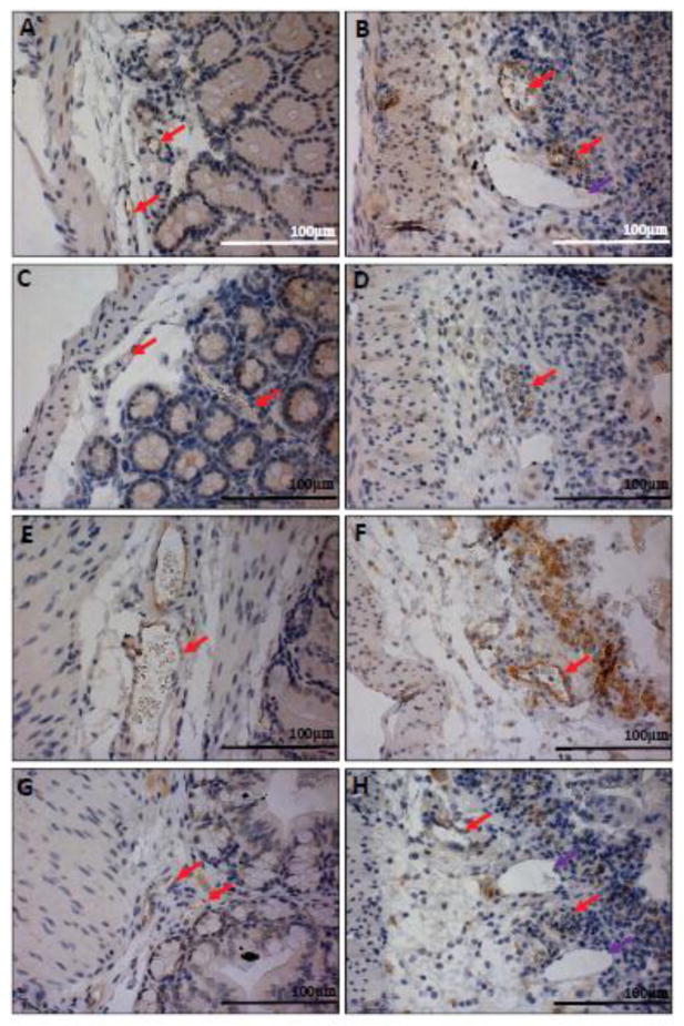
Fig. 10.
Immunohistochemical analysis of cecum adhesion molecule expression in ECs. In addition to the colon, cecum tissues were examined for adhesion molecule expression between WT and GPR4 KO mice. Similar to colon, GPR4 KO-DSS mice had a reduction in the expression of E-selectin and VCAM-1 in ECs. E-selectin protein expression could be visualized as brown signals in (A) WT-control, (B) WT-DSS, (C) GPR4 KO-control, and GPR4 KO-DSS mucosal blood vessels. VCAM-1 protein expression could be visualized in (E) WT-control, (F) WT-DSS, (G) GPR4 KO-control, and (H) GPR4 KO-DSS mucosal blood vessels. 40× microscope objective. Red Arrows indicate blood vessels.

