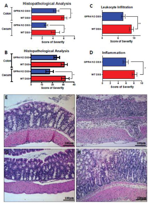
Fig. 2.
Histopathological analysis of mouse colon and cecum. Histological features of colitis were examined to further assess the degree of disease activity in the mice by veterinary and human pathologists using complementary, yet distinct scoring systems. Overall, GPR4 KO DSS mice had reduced histopathological scores in cecum and colon when compared to WT DSS mice. (A) Veterinary pathologist and (B, C, D) human pathologist assessment of colon and cecum. (C) Reduced leukocyte infiltration was observed spanning from the cecum to distal colon in GPR4 KO DSS mice compared to WT DSS mice. (D) Overall inflammation was reduced in GPR4 KO DSS mice compared to WT DSS mice in tissues spanning from the cecum to distal colon. Representative H&E staining pictures of colon in (E) WT control, (F) WT-DSS, (G) GPR4 KO control, and (H) GPR4 KO DSS using a 20× microscope objective. Data are presented as mean ± SEM and analyzed for statistical significance between WT-DSS and GPR4 KO DSS groups using the unpaired t-test. WT-Control (n=12), WT-DSS (n=13), GPR4 KO Untreated (n=12), and GPR4 KO DSS (n=18) tissues were used for analysis. (*P < 0.05, **P < 0.01)

