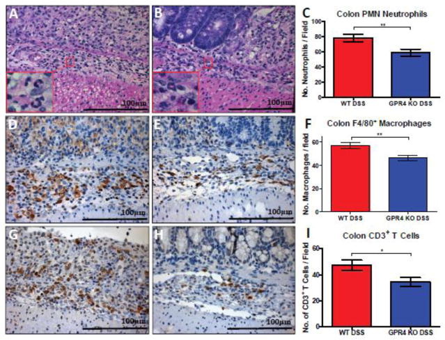
Fig. 3.
Immune cell infiltrate quantification in colon mucosa. GPR4 KO-DSS mice (n= 4–5) had reduced numbers of neutrophils, macrophages, and T cells in the mucosa of the colon compared to WT-DSS mice (n= 4–5). (Fig. 3A–C) Neutrophil quantification based on polymorphonuclear (PMN) morphology and cytoplasmic staining, (Fig. 3D–F) F4/80+ macrophages, and (Fig. 3G–I) CD3+ T cells. 40× microscope objectives. Statistical analysis was performed using the unpaired t-test between WT-DSS and GPR4 KO-DSS groups. (*P < 0.05, **P < 0.01)

