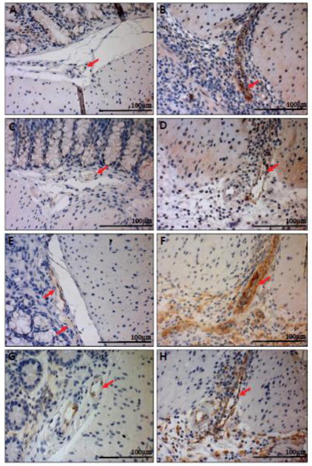
Fig. 9.
Immunohistochemical analysis of E-selectin and VCAM-1 protein expression in mouse colon tissues. As whole tissues are not ideal for analyzing endothelial cell specific gene expression, we performed IHC to analyze adhesion molecules E-selectin and VCAM-1 protein expression in ECs within the tissue. GPR4 KO-DSS mice have reduced E-selectin and VCAM-1 protein expression in colonic mucosal vasculature when compared to WT-DSS mice. E-selectin expression could be visualized as brown signals in (A) WT-control, (B) WT-DSS, (C) GPR4 KO-control, and (D) GPR4 KO-DSS colon tissues. VCAM-1 expression could be visualized in (E) WT-control, (F) WT-DSS, (G) GPR4 KO-control, and (H) GPR4 KO-DSS colon tissues. 40× microscope objective. Red Arrows indicate blood vessels.

