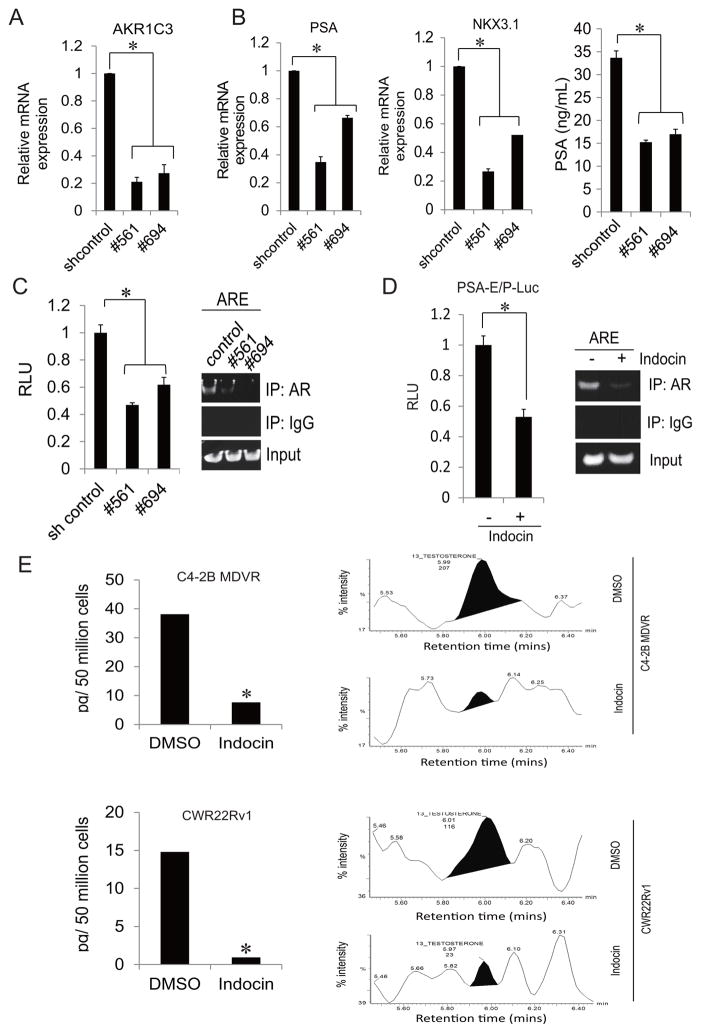
Figure 2. AKR1C3 inhibition suppressed testosterone level and AR activity in prostate cancer.
A. C4-2B MDVR cells were transiently transfected with control shRNA or AKR1C3 shRNA (#561, #694) for 2 days. Total RNA was extracted and AKR1C3 mRNA level was examined by qRT-PCR. B. C4-2B MDVR cells were transiently transfected with control shRNA or AKR1C3 shRNA (#561, #694), Total RNA was extracted, PSA and NKX3.1 mRNA levels were examined by qRT-PCR, and the supernatants were collected and subjected to PSA ELISA. C. C4-2B MDVR cells were transiently transfected with control shRNA or AKR1C3 shRNA (#561, #694) with PSA-E/P-luc reporter. The luciferase activity was detected by dual luciferase reporter system (left) and whole cell lysates were subjected to ChIP assay (right). D. C4-2B MDVR cells were transiently transfected with PSA-E/P-luc reporter, followed by treatment with DMSO or 20 μM Indocin for 3 days. Luciferase activity was detected by dual luciferase reporter system (left) and whole cell lysates were subjected to ChIP assay (right). E. C4-2B MDVR (top) and CWR22Rv1 (bottom) cells were treated with DMSO or 20 μM Indocin in serum free, phenol red free RPMI1640 medium for 3 days. Fifty million cells were collected per treatment and testosterone level was examined by LC-MS. * p<0.05. Indocin: Indomethacin.

