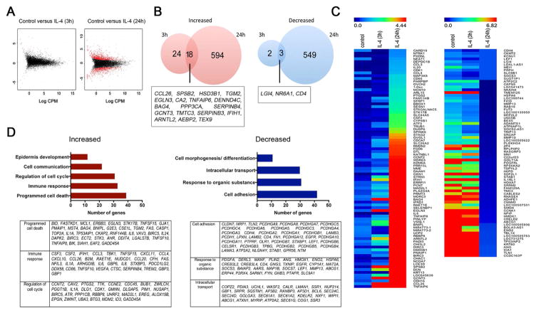
Figure 1. IL-4 modifies human keratinocyte (HK) gene expression.
HK were differentiated for 2 days with calcium chloride and stimulated with IL-4 for 3 and 24 hours before RNA-seq analysis. (A) Graphical representation of genes that were enriched at least 2-fold in IL-4-stimulated keratinocytes (red dots). (B) Venn diagram representing the number of genes altered by IL-4 after 3 and 24 hours. (C) Heatmap comparison of gene enrichment in IL-4-stimulated HK using MeV software. (D) Gene ontology analysis using the differential expression of genes at 24 hours after IL-4. Clustering was performed using David bioinformatics database analysis. Data represent average of two biological replicates for each condition. *p<0.05 and FDR<0.1

