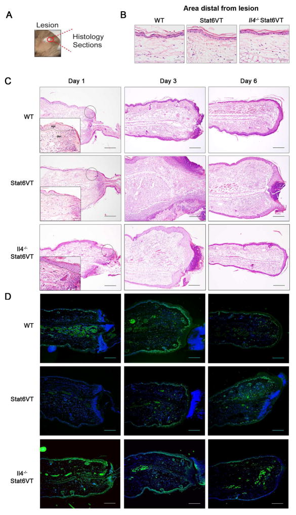
Figure 4. Stat6VT mice exhibit delayed wounding healing.
Lesions were monitored by H&E in ears of WT, Stat6VT and Il4−/− Stat6VT mice at days 1, 3 and 6 after wounding (dermis (der) and epidermis (epi) separated by dashed lines). (A) Representation of ear punch. (B) Visualization of histological sections from distal areas from lesion and (C) lesion re-epithelialization and wounding closure. (D) Immunofluorescence for the proliferation marker Ki67 (green fluorescence) in the ears of WT, STAT6VT and Il4−/− Stat6VT mice in indicated days after punch. Nuclei are stained with DAPI in blue. Scale bar: 200μm. Pictures are representative of 3–4 mice from 2 independent experiments.

