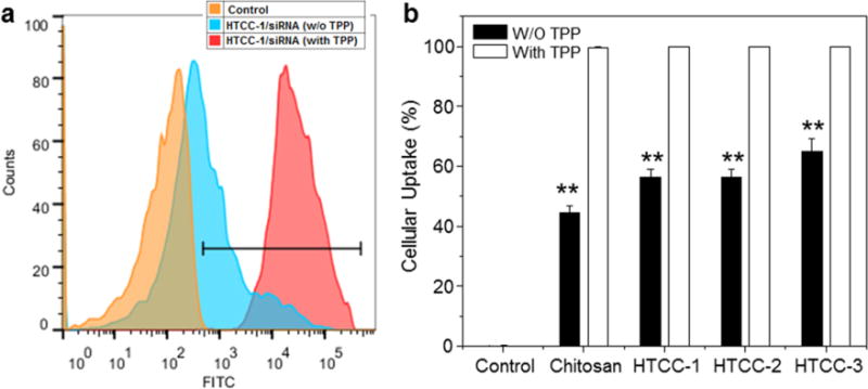
Fig. 6.

Quantification of cellular uptake of various NPs (weight ratio, 60:1) by Raw 264.7 macrophages. (a) Representative flow cytometry histograms of fluorescence intensity for cells treated with TPP-HTCC-1/siRNA NPs for 5 h. (b) Percentage of FITC fluorescence-positive cells after treatment with various NPs for 5 h. FITC-siRNA (100 nM) was used for the transfection. Each point represents the mean ± S.E.M. (n = 3; *P< 0.05 and **P< 0.01, Student’s t-test).
