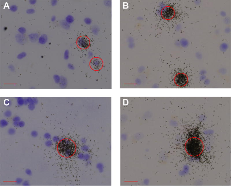
Figure 2. Patterns of SST-positive grain clusters in amygdala.

Circles with a 15μm diameter (A–B) or 25μm (C–D) were centered over SST-positive nuclei. Circles were centered over SST-positive nuclei and other neurons in every counting frame, and the ratio of area covered by silver grains was calculated in the corresponding darkfield image. Compared to the small clusters (A and B), large clusters (C and D) showed considerable grain overlap and higher signal intensity, making it more difficult to count the number of individual grains in large clusters. Thus, we instead used the ratio of area covered by grains within each circle as a proxy measure of amount of SST per positive cell (both small and large). Scale bar = 15μm.
