Figure 3.
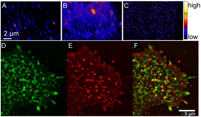
High-resolution SIMS and complementary immunofluorescence imaging shows hemagglutinin does not cluster in plasma membrane domains that are enriched with cholesterol and sphingolipids. High-resolution SIMS images of a region on a mouse fibroblast cell that stably expressed influenza hemagglutinin (Clone 15 cell line). (A) High-resolution SIMS image of the 19F− counts shows the distribution of immunolabeled hemagglutinin in the plasma membrane. Comparison to the (B) 15N-enrichment and (C) 18O-enrichment images that were simultaneously acquired indicates hemagglutinin is not located in cholesterol- and sphingolipid-enriched domains. Reprinted from Wilson et al. (2015). Copyright (2015) with permission from Elsevier. Total internal reflectance microscopy detection of (D) BODIPY-sphingolipids (green) and (E) hemagglutinin (red) in the plasma membrane of a living Clone 15 cell. (F) Overlay shows little colocalization between the sphingolipids and hemagglutinin (yellow). Scale bar is 5 μm. Reproduced with permission from Frisz et al. (2013b). Copyright 2013 National Academy of Sciences, U.S.A.

