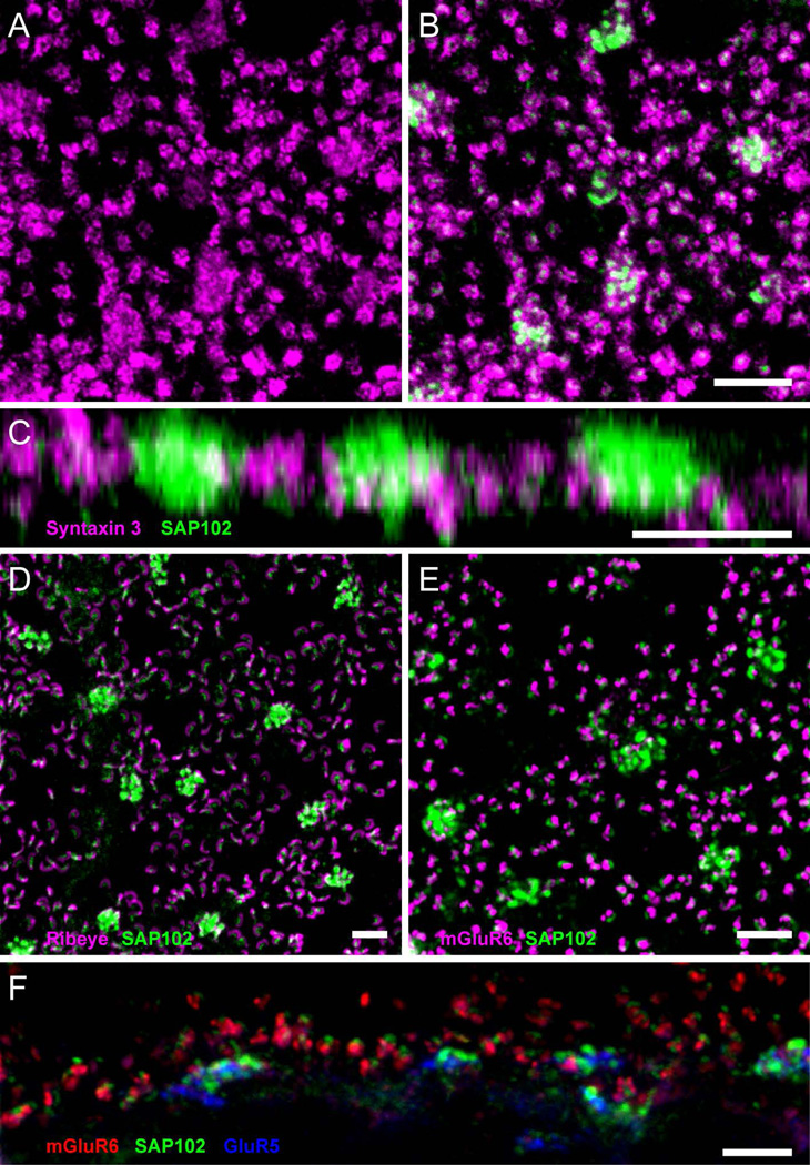
Figure 3. Association of SAP102 with synaptic elements in the OPL.
A, Wholemount immunofluorescence labeling in the OPL of syntaxin 3 (magenta), revealing small clusters labeling synaptic terminals of rods, and diffuse cellular staining in cone terminals. B, Single optical section through the OPL double labeled for SAP102 (green) and syntaxin 3 (magenta). SAP102 labeling appears in gaps in syntaxin 3 labeling. C, Z-axis projection of a series of optical sections taken at 0.35 µm intervals through the OPL. SAP102 puncta in clusters were localized slightly above the cone plasma membrane labeled with syntaxin 3, suggesting that SAP102 is inside of cone pedicles and not in their plasma membrane. One punctum was typically found per rod spherule. D, Wholemount immunofluorescence labeling in the OPL of SAP102 (green) and the synaptic ribbon protein ribeye (magenta). E, Wholemount immunofluorescence labeling in the OPL of SAP102 (green) and mGluR6 (magenta), labeling the tips of ON bipolar cell dendrites. F, Vertical section through the OPL with labeling of SAP102 (green), mGluR6 (red) and GluR5 (blue). The SAP102-IR puncta were always located above ON bipolar cell dendrites (mGluR6) in rods and slightly above OFF bipolar cell basal contacts (GluR5) in cones. Scale bars: 5 µm.

