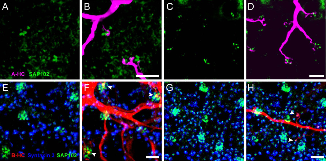
Figure 4. SAP102 is located in B-type horizontal cells.
(A–D) Distribution of SAP102 (green) immunoreactivity relative to a Neurobiotin-injected A-type HC (magenta) in the OPL. A and C: Distribution of SAP102 immunoreactivity in the OPL. B and D: Stacks from 15 single optical sections at 0.25 µm increments are shown. A-type HC dendrites labeled with Neurobiotin were closely associated with clusters of SAP102 immunoreactive puncta in cones, but not co-localized. (E–H): Distribution of SAP102 immunoreactivity (green) relative to a Neurobiotin-injected B-type HC (red) in the OPL. E and G: Distribution of SAP102 immunoreactivity in the OPL. The location of photoreceptor terminals is shown with syntaxin 3 labeling (blue). Stacks from 17 single optical sections at 0.25 µm increments are shown. F and H, Relationships of B-type HC dendrites (F) and axon terminals (H) with SAP102. Both B-type HC dendrites (F) and axon terminals (H) colocalized with SAP102 (arrowheads) at contacts with cone terminals and rod spherules respectively. Scale bars: 5 µm.

