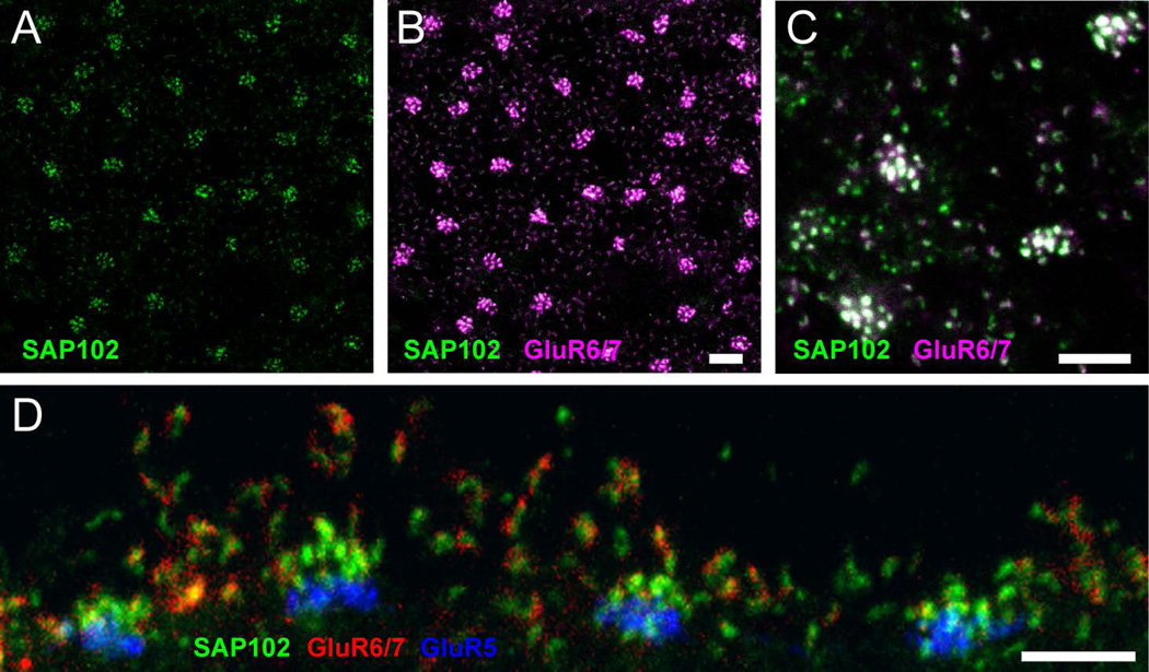
Figure 5. Association of SAP102 with kainate receptor GluR6/7.
(A–C), Wholemount immunofluorescence labeling in the OPL of SAP102 (green; alone in A) and GluR6/7 (magenta). B, A stack from 6 single optical sections at 0.35 µm increments is shown. GluR6/7-IR appeared as discrete puncta colocalized with SAP102. C, Single optical section confirming that GluR6/7 colocalization with SAP102 was not due to projection of labels at different depths. D, Vertical section through the OPL labeled for SAP102 (green), GluR6/7 (red) and GluR5 (blue). GluR6/7 labeling was clearly colocalized with the clusters of SAP102 associated with the invaginations of rod spherules. Scale bars: 5 µm.

