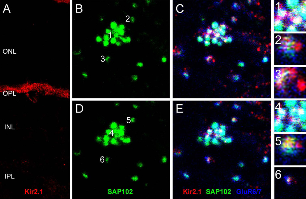
Figure 6. Association of SAP102 with inward-rectifier potassium channel Kir2.1.
A, Vertical section through the retina labeled for Kir2.1. Kir2.1 exhibited a mixture of diffuse and punctate patterns in the OPL and surrounded the somas of HCs. (B–E), Wholemount immunofluorescence labeling in the OPL of SAP102 (green), Kir2.1 (red) and GluR6/7 (blue) in two consecutive optical sections. B and C represent a single optical section in the lower portion of the OPL separated by 0.35 µm from a higher section in the OPL represented in D and E. B and D, SAP102 alone. C and E, Kir2.1-IR (red) was diffusely present below SAP102 clusters, but puncta were colocalized with SAP102 at the tips of HC processes. Numbers in B and D indicate individual SAP102 puncta at contacts with cone (1, 4) and rod (2–3, 5–6) terminals that are shown in the insets. Kir2.1 colocalizes with SAP102 at the level of the cones (inset 1) and at the level of the rods (insets 5 and 6). Scale bar: 5 µm.

