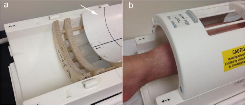
Fig. 1.

(a) Lower part of the 3T sodium knee coil. Inside the coil there are four calibration standards fixed in a phantom holder. A concave cover with a hard smooth surface (as pointed out by the white arrow) is slid open to display the phantoms. (b) During imaging, the skin is in direct contact with the surface of the cover.
