
FIGURE 3.
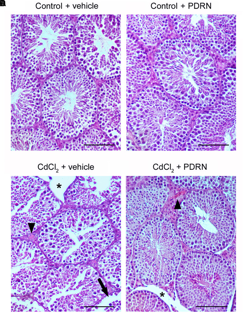
Representative histological sections of a testis from a control plus vehicle animal (A), a control plus PDRN animal (B), a CdCl2 (2 mg/kg i.p.) plus vehicle animal (C), and a CdCl2 (2 mg/kg i.p.) plus PDRN (8 mg/kg i.p.) animal (D). (A,B) Seminiferous tubules and extratubular compartment show normal morphology. (C) Arrow, Round spermatids and spermatogonia detached from the basal membrane; ∗, marked edema of the extratubular compartment; Arrowhead, hemorrhagic extravasation. (D) ∗, mild interstitial edema; Arrowhead, enlarged vessels in the extratubular compartment. (Scale bar: 50 μm).