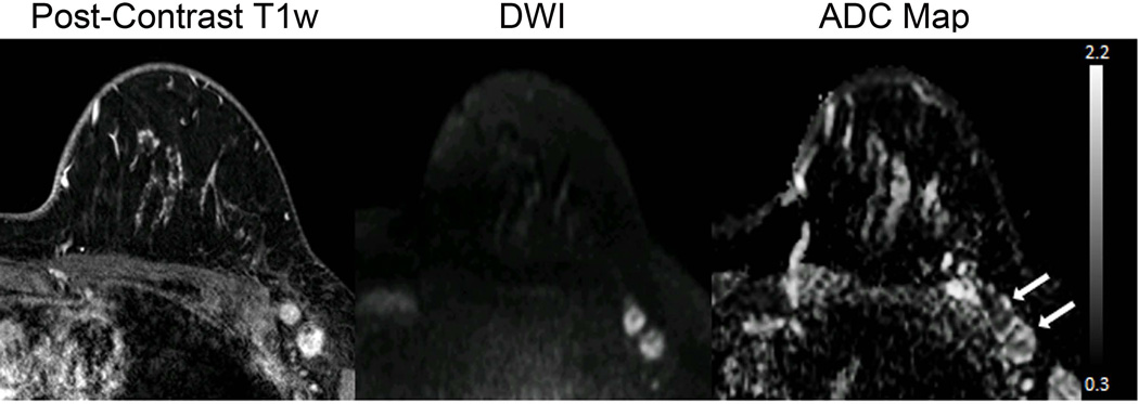
Figure 6.
Axillary metastasis. A 31-year-old woman underwent staging MR imaging after newly diagnosed invasive ductal carcinoma in the left breast. Multiple abnormal level 1 axillary lymph nodes with biopsy proven axillary node metastasis are present in the left axilla, seen as enhancement on the post-contrast T1-weighted image and as hyperintense on the DW (b = 1000 s/mm2) image due to restricted diffusion. The ADC map demonstrates corresponding low values, with ADC = 0.83 ×10⁻³ mm2/s and ADC = 1.17 ×10⁻³ mm2/s for the anterior and posterior nodes, respectively (arrows).

