Fig. 5.
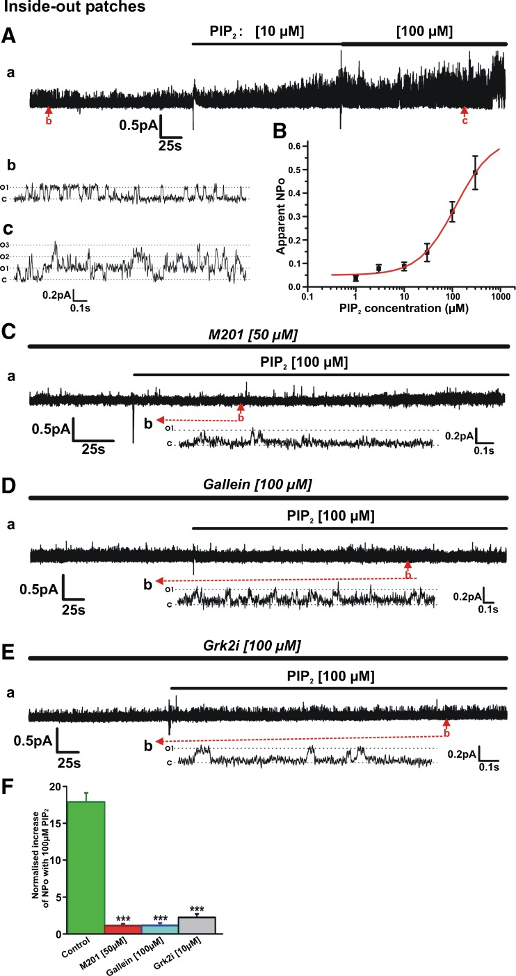
Gβγ inhibition prevents activation of Kv7.4 channels by exogenous PIP2. A Representative inside-out patch recording showing stimulatory action of PIP2. Subpanels b and c are 1.75-s segments of channel openings in the absence (b) and presence of PIP2 (100 μM, c) taken from long-term recording (a). Closed state and multiple open states are denoted by C and O1–O3. B Mean concentration-effect for PIP2 (n = 4–11). C Representative inside-out patch recording showing that in the continued presence of 50 μM M201 (continuation of recording from patch shown in Fig. 4D), 100 μM PIP2 applied to the patch failed to activate the channels. Subpanel b is a 1.5-s segment of channel openings taken from long-term recording (a). Closed state and multiple open states are denoted by C and O1. D Representative inside-out patch recording showing that in the continued presence of 100 μM gallein, 100 μM PIP2 applied to the patch failed to activate the channels. Subpanel b is a 2.5-s segment of channel openings taken from long-term recording (a). E Representative inside-out patch recording showing that in the continued presence of 100 μM Grk2i (continuation of recording from patch shown in Fig. 4E), 100 μM PIP2 applied to the patch produced only negligible activation of the channels. Subpanel b is a 2.8-s segment of channel openings taken from long-term recording (a). F Normalized increase in NPo produced by 100 μM PIP2 applied to inside-out patches in the absence and presence of different Gβγ inhibitors (n = 4–6)

